USER MANUAL | ICOS | INSTRUCTIONS | UM/ICOS-EN REV. B.2
35
Spectrum Display
Click the Display button on the
User Interface Control Bar to switch to Spectrum Display.
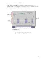
The top plot shows the voltage from the photo-detector as the laser scans across the
absorption features.
The bottom plot shows the corresponding optical absorption displayed as black circles,
and the peak fit resulting from signal analysis as a blue line.
Figure 19 and Figure 20 show the
Spectrum Displays for the following analyzers:
GLA151-N2OM1
GLA151-N2OCM
GLA151-N2OM1 Portable Methane / Nitrous Oxide Analyzer
The measured N
2
O concentration is shown in parts per billion (ppb), and CH
4
and H
2
O in
parts per million (ppm) on the bottom of the
Spectrum Display.
Figure 19: Spectrum Display (GLA151-N2OM1)

