49
VIDEO
MICROSCOPE
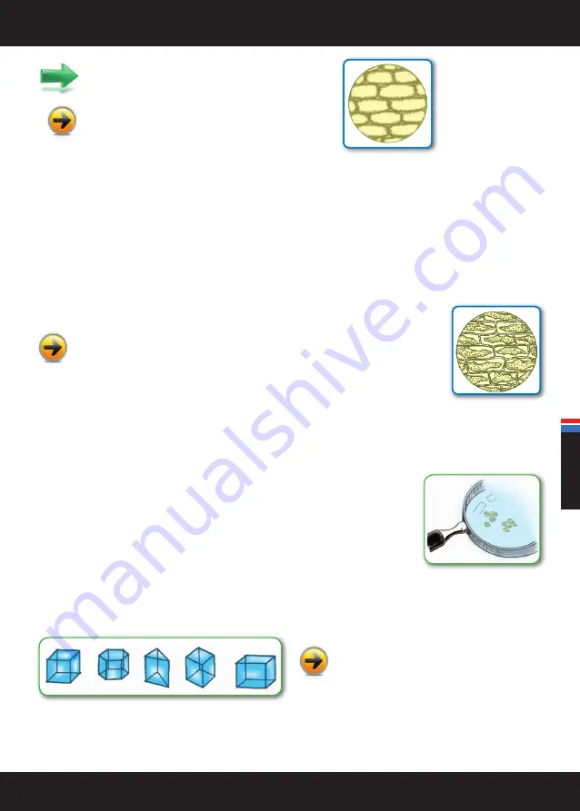
Pay close attention:
you should be able to see a series of
rectangles tied together. These are the skin cells.
Inside the cell you could see also a round shape:
it is
the nucleus, which you will also be able to detect in the
following observations.
EPIDERMAL CELLS OF AN ONION WITH SALT WATER
1)
First prepare a solution of table salt. Put a few teaspoons of salt into a glass of water.
One solution is a mixture in which two or more substances can no longer be distinguished. Normally when we talk of solutions we mean
mixtures in the liquid state, constituted by a liquid solvent containing the dissolved solute.
2)
Prepare a slide with the onion epidermis as described in the observation above.
3)
Use a plastic pipette to place two drops of the table salt solution on the skin sample. Place a piece of clear plastic on top: the
coverslip.
After a while (10/15 minutes), observe the skin with the video microscope. What difference do
you see compared to the previous observation? In a plant cell, the cell membrane is detached
from the wall because the salty water that surrounds it draws water from the inside. This phenomenon is
called
PLASMOLYSIS
.
Note:
Now if you put a few drops of distilled water (ask an adult: it is the water that is used for ironing and car batteries) on the
skin sample you’ve just observed, you can see the opposite phenomenon. The cells inflate with distilled water that enters by
osmosis into them because the internal fluid is richer in salts.
SALT AND SAND CRYSTALS
Sand
Find some sand. Distribute it on an observation surface and grab your video camera. Pay close
attention. You will note the shape of the grains of sand. There are two types of grains: the grains
with sharp edges from young beaches or short rivers, and the rounded edged grains from ancient
beaches or those that have been transported by very long rivers.
Crystals
Crystals are a solid substance whose particles (atoms and ions) are arranged in a geometric and
repetitive pattern.
Observe salt crystals. Use a half tablespoon of table
salt (you can use fine grains or the larger course salt)
and arrange them on an observation surface.
Adjust the focus by turning the ring and take in the shapes of the crystals.
sand grains seen with a magnifying
glass: with sharp edges and
rounded edges
ENGLISH
Summary of Contents for VIDEOMICROSCOPE
Page 52: ...52 VIDEOMICROSCOPE ANTECKNINGAR MERKNADER DINE EGNE NOTER MUISTIINPANOJA NOTES...
Page 53: ...53 VIDEOMICROSCOPE ANTECKNINGAR MERKNADER DINE EGNE NOTER MUISTIINPANOJA NOTES...
Page 54: ...54 VIDEOMICROSCOPE ANTECKNINGAR MERKNADER DINE EGNE NOTER MUISTIINPANOJA NOTES...
Page 55: ...55 VIDEOMICROSCOPE ANTECKNINGAR MERKNADER DINE EGNE NOTER MUISTIINPANOJA NOTES...
Page 56: ...56 VIDEOMICROSCOPE ANTECKNINGAR MERKNADER DINE EGNE NOTER MUISTIINPANOJA NOTES...
Page 57: ...57 VIDEOMICROSCOPE ANTECKNINGAR MERKNADER DINE EGNE NOTER MUISTIINPANOJA NOTES...
Page 58: ...58 VIDEOMICROSCOPE ANTECKNINGAR MERKNADER DINE EGNE NOTER MUISTIINPANOJA NOTES...
Page 59: ...59 VIDEOMICROSCOPE ANTECKNINGAR MERKNADER DINE EGNE NOTER MUISTIINPANOJA NOTES...
Page 60: ...60 VIDEOMICROSCOPE ANTECKNINGAR MERKNADER DINE EGNE NOTER MUISTIINPANOJA NOTES...
Page 61: ...61 VIDEOMICROSCOPE ANTECKNINGAR MERKNADER DINE EGNE NOTER MUISTIINPANOJA NOTES...
Page 62: ...62 VIDEOMICROSCOPE ANTECKNINGAR MERKNADER DINE EGNE NOTER MUISTIINPANOJA NOTES...
Page 63: ...63 VIDEOMICROSCOPE ANTECKNINGAR MERKNADER DINE EGNE NOTER MUISTIINPANOJA NOTES...


