24
•
Imaging System Hardware
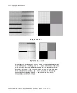
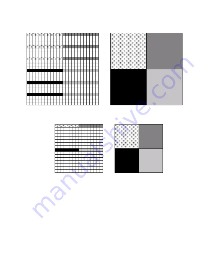
D) 60-µm Data Scan
E) Preview Scan (40-µm)
Representation of how the GenePix scanner samples 5-µm spots and averages them
to calculate the values of the image pixels for the various scan modes and pixel sizes.
On the left side of each image pair is the raw data scan from the GenePix Scanner.
Each square represents a 5 µm × 5 µm spot as scanned by the 5-µm laser beam.
On the right are the resultant image pixels at various resolutions. The shading
indicates the spots that are scanned by the GenePix scanner as well as the
corresponding pixels in the final image.
GenePix 4200A User’s Guide, Copyright 2005 Axon Instruments / Molecular Devices Corp.


