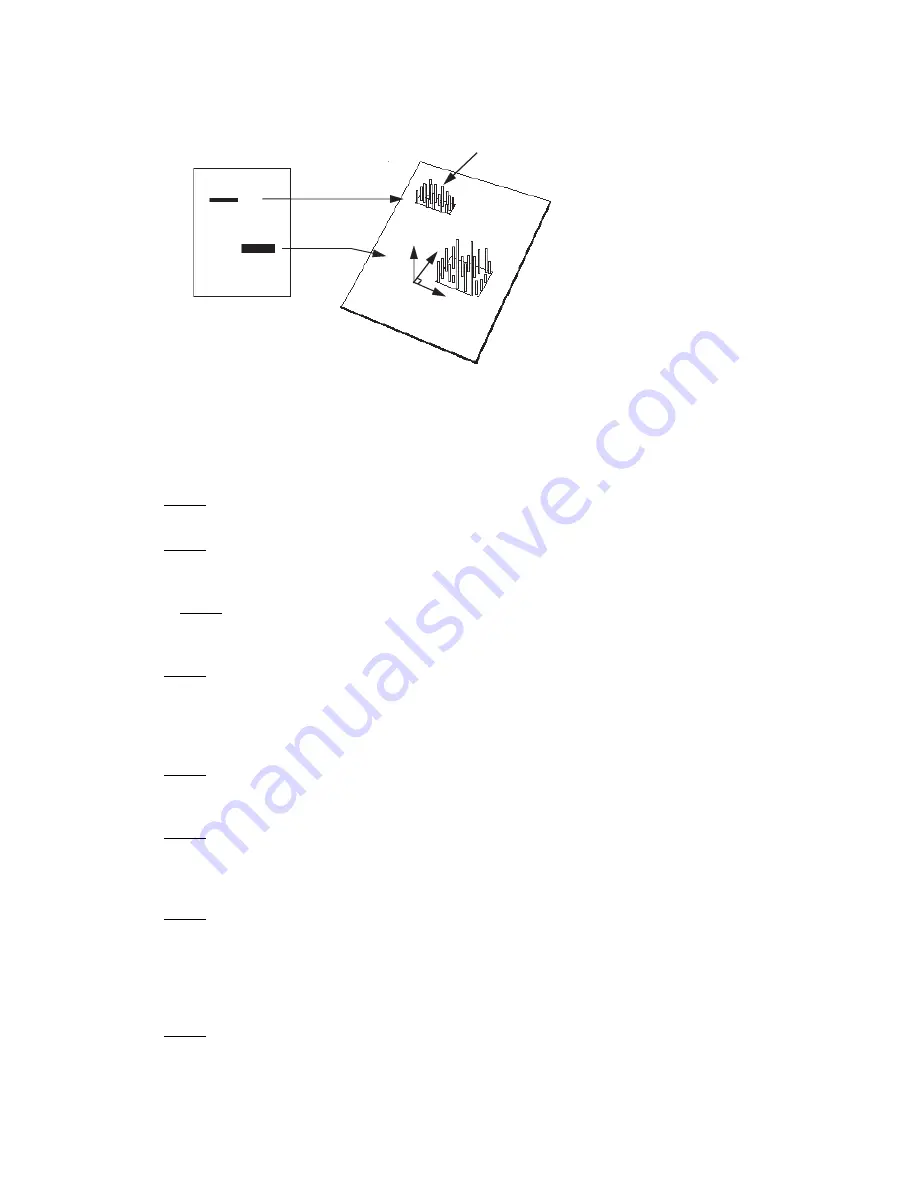
Fig. 2.8. Two and three-dimensional representations of a digitized image.
2.4 Overview of the Imaging Process
2.4.1 Steps in Fluorescence Imaging
The procedure for imaging fluorescent samples has four steps.
Step 1 involves thoroughly cleaning the sample tray with ethanol, using a clean soft cloth or
lint free paper towel.
Step 2 involves placement of the sample onto the tray. Several accessories, such as gel
holders, spacers and microtiter plate inserts are provided to optimize position of your sample
on the tray.
In Step 3 the tray with sample loaded is pushed into the laser scanner and the sample
imaged by direct fluorescence excitation. The host computer builds a digitized image of the
sample by tracking each pixel's signal as the scanner head moves over the screen surface.
Step 4 Once the sample image is collected, it can then be reviewed and analyzed using an
appropriate software package.
2.4.2 Steps in Storage Phosphorescence Imaging
Storage phosphor imaging is a simple four-part process. (Figure 2.9)
Step 1 involves erasing the reusable phosphor screen to remove any background or
residual image. This normally takes 10 minutes. The screens should be erased to the
background level of 100 counts or less.
Step 2 in the process involves placement of the prepared sample in an exposure cassette
for subsequent close proximity exposure to the imaging screen. The captured signal
generates a latent image of the sample, which is encoded in the number and pattern of
charged phosphor crystals.
Step 3 involves placing the screen in a laser scanner. As each pixel of the screen is
scanned, the electrons in charged areas of the latent image return to the ground state,
releasing energy in the form of emitted photons of visible light. The emitted photons are
collected and precisely counted by a photomultiplier tube, generating an intensity for each
scanned pixel. This intensity is expressed in counts or pixel density units, which are
analogous to the optical density of exposed x-ray film in autoradiography.
Step 4 is analogous to the procedure for imaging fluorescence samples, the resulting
image can then be reviewed and analyzed using an appropriate software package.
2-D view
3-D vie
w
Intensity
Single Pixel
10


