Manual, Ultra High Sensitivity Aerosol Spectrometer (UHSAS)
DOC-0210 Rev E-4
1 4
© 2017 DROPLET MEASUREMENT TECHNOLOGIES
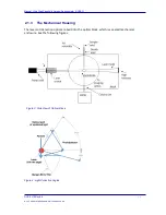
Figure 3 and Figure 4 show two views of the optical cavity. Figure 3 shows the general layout of
the aerosol inlet, placement of the solid state laser and particle stream with respect to the
mangin mirror pair.
Figure 4 shows the optical path of light collected from scattering by particles that pass through
the laser. The detector is located at 90
0
to the path of the particle through the laser. (The laser
can be envisioned as shining into the page.) The mangin mirror pair collects the scattered light
over solid angles of 90
0
± 57
0
(33
0
– 147
0
), excluding the angles subtended by the circular
opening of the mangin mirrors 90
0
± 14.8
0
(75.2
0
– 104.8
0
).
For the purpose of calculating scattering cross sections with Mie scattering, the collection angles
are 33
0
-75.2
0
and 104.8
0
-147
0
.
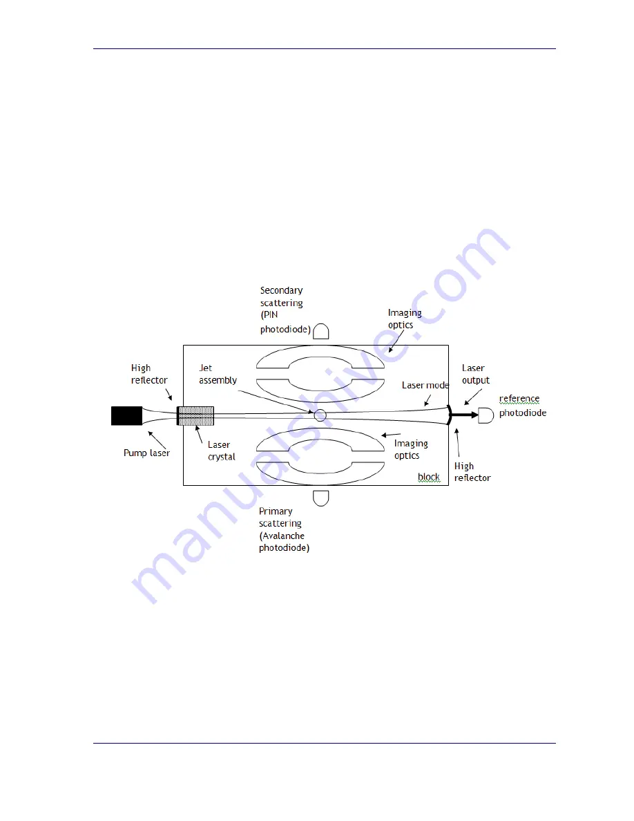
Figure 5 shows a top view of the optical block.
Figure 5: Top View of Optical Block

