8
Section 2. Operation
Scanning with Doppler Signals
When setting up for Doppler signal imaging:
•
A linear array transducer performs best for beam steering tests.
•
Use a small sample volume (gate) placed as closely as possible to the middle of the vessel.
Perform the scan:
1
Using the same scanning plane orientation used in
Scanning for Color Flow Imaging
place transducer on phantom.
2
On ultrasound system, select
B-mode
display and visualize the horizontal vessel.
3
Activate ultrasound system’s
PW Doppler (
Pulsed-Wave Doppler) mode.
4
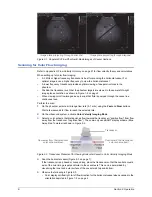
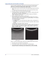
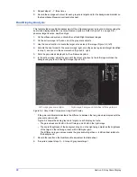
Generate a spectral Doppler signal with beam steered towards the right, as shown in Figure 2-
5, left image.
5
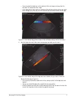
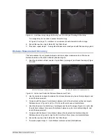
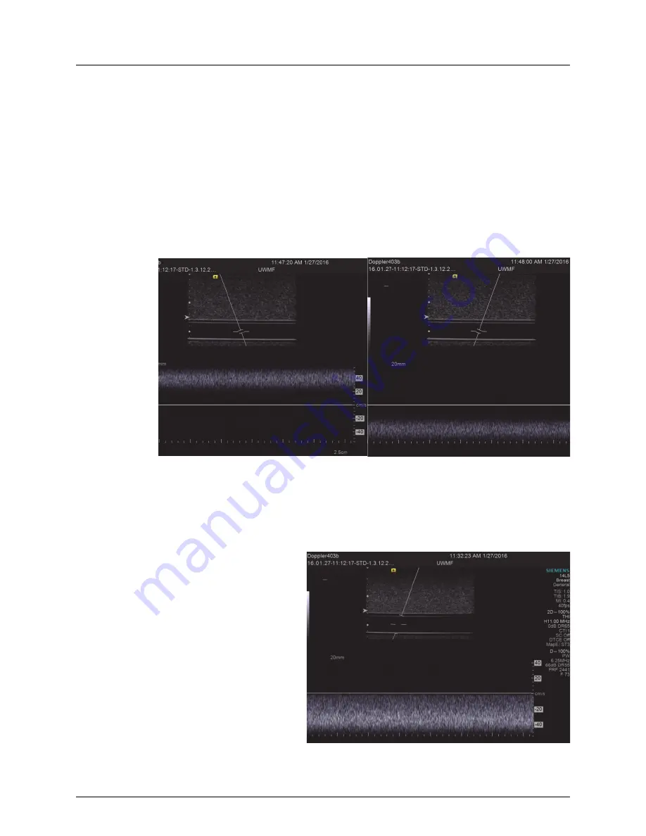
Without moving transducer, steer Doppler beam towards the left and generate another spectral
Doppler signal, as shown in Figure 2-6.
Figure 2-5. Doppler Spectral Display of Horizontal Vessel, Beam Steering, Continuous Flow
Observe the following in Figure 2-5:
• Flow direction as established in Figure 2-2 on page 6 is verified, and flow is continuous.
• Vessel contents appear black in the gray scale images, verifying the low echogenicity of the
blood-mimicking fluid.
6
Increase sample volume so
gated region includes entire
diameter of vessel.
Observe the following in
Figure 2-6:
• Flow direction is away
from the transducer.
• Compared with Figure 2-
5, a wider range of flow
velocities is displayed,
from maximum along the
vessel axis to near zero at
the vessel walls.
• A parabolic flow profile is
seen in this part of the
vessel, using a mid-range
flow rate.
Flow towards transducer
Flow away from transducer
Figure 2-6. Doppler Spectral Display of Horizontal Vessel,
Continuous Flow, Full Gated Region

