Harmonic Imaging in Medical Ultrasound
33
Appendix B: Harmonic Imaging
Harmonic Imaging in Medical Ultrasound
Harmonic imaging has become an important signal processing and display mode in medical
ultrasound. In harmonic imaging an ultrasound pulse is sent from the transducer at a nominal
(fundamental) frequency. If the pulse has a high enough amplitude, it undergoes a nonlinear
distortion as it travels through tissues. This results in the creation of harmonic components of the
original pulse frequency. The creation of harmonics is a gradual process, peaking at a depth that
depends on the medium and original pulsing conditions, and then tapering off with increasing depth.
Any detected echoes consist of both fundamental and harmonic frequencies.
When harmonic imaging is activated, the echo signal’s fundamental frequency components are
filtered out, or at least subdued, and the harmonic components are emphasized. In most cases, the
second harmonic, i.e., a signal whose frequency is twice that of the fundamental frequency, is
detected and displayed. The result is that better image quality can often be attained for a given depth
than if the reception had been at the fundamental frequency.
One way in which image quality is improved using harmonics is through the reduction of
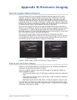
reverberations and unwanted clutter. Figure B-1, left, shows a typical image in which reverberation
echo signals are seen within the common carotid artery, as indicated by the white arrow. These
reverberations are reduced when harmonic imaging is activated (right).
Figure B-1. B-Mode Images of Common Carotid Artery, Imaging Comparison
Phantom Use in Harmonic Imaging
Phantoms can help illustrate differences in quality between images obtained using fundamental
imaging and images obtained using harmonic imaging.
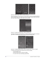
Figure B-2 on page 34 illustrates a comparison among images of the hypoechoic spheres in a
Sono408 (Model 408) phantom.
• Left image was generated using a 14 MHz frequency transducer operating in fundamental
mode.
• Center image was generated using a lower frequency (7 MHz, L7) transducer operating in
fundamental mode at 6 MHz.
• Right image was generated at 7 MHz, with an L7 transducer operating in harmonic mode.
The L7 transducer was operating in fundamental imaging mode at 6 MHz to generate the middle
image and in harmonic mode to generate the image on the right. The hypoechoic structures appear
to be sharpest in the image on the right.
Fundamental Imaging
Harmonic Imaging


