Directional Discrimination
11
1
Set the phantom to CONTINUOUS mode with a low (1–2 ml/s) flow rate.
2
Record the mode and rate.
3
On the ultrasound system, set the color flow output power and color gain for maximum
sensitivity without excessive noise.
4
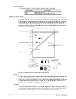
Scan the diagonal vessel.
5
Toggle between color flow and B-mode.
a.
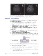
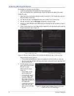
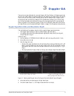
Observe whether the color flow information is displayed only within the vessel.
b.
Record scanner settings that produce poor correlation.
c.
Note whether the highest flow rate occurs at the vessel’s center and the lowest flow rate at
the vessel wall.
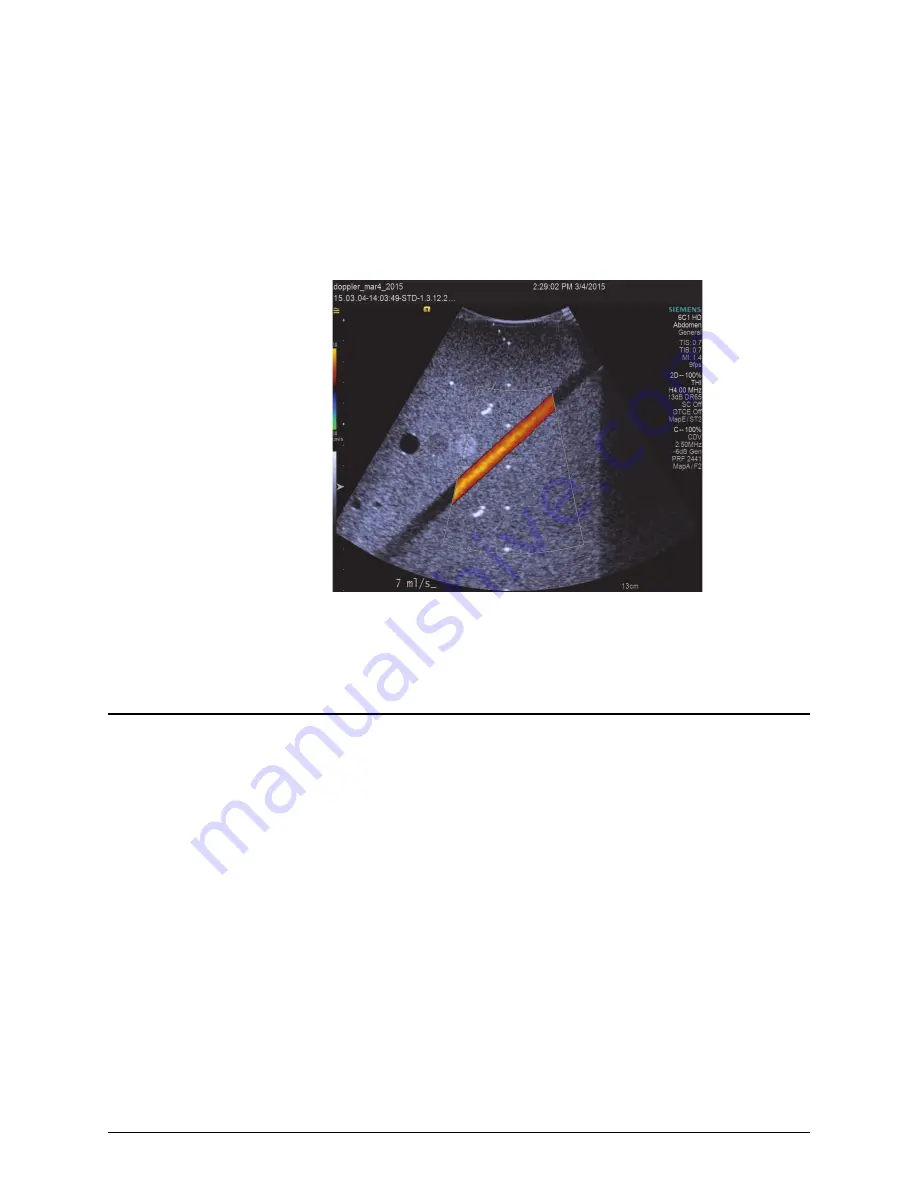
Figure 3-3. Scanning the Diagonal Vessel for Color Flow and B-Mode Image Correlation
6
Record the results on the Doppler QC Test Data sheet.
7
If required, repeat steps
at a range of flow rates from low to high.
Directional Discrimination
The Directional Discrimination test evaluates the scanner’s ability to accurately display flow
direction.
With the beam perpendicular to the flow, Doppler signals and the spectral broadening artifact should
be equally positive and negative, and the magnitude of the signals should be the same.
Perform the test:
1
Set the phantom to CONTINUOUS mode and set the flow rate to produce a laminar flow, at a
flow rate low enough that aliasing does not occur.
2
Record the mode and rate.
3
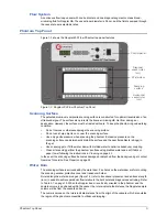
Scan the horizontal vessel.
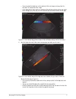
Place the transducer so that the beam is perpendicular to the vessel axis (Figure 3-4 on page
12).
The spectral display should be equally positive and negative, mirroring across the baseline.

