GE M
EDICAL
S
YSTEMS
D
IRECTION
2392751-100, R
EVISION
3
V
IVID
™ 4 S
ERVICE
M
ANUAL
4-12
Section 4-4 - Image Testing: 2D/M/CFM/Doppler
4-4-1-3
2D Penetration Test
4-4-1-4
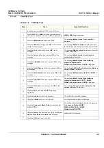
CFM Noise Floor Test
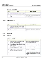
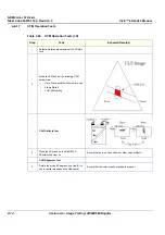
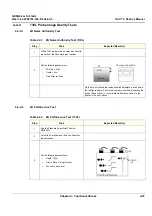
Table 4-20 2D Noise Penetration Test (3S)
Step
Task
Expected Result(s)
1
Use the Standard Imaging Phantom
RMI403GS.
2
Select a cardiac preset.
3
Set the following parameters, and scan the
phantom at position (3):
•
Gain: 37
•
Power: 0 dB
•
Depth: 20cm
•
Focus: Max depth
•
Dynamic Range: 65dB
4
Record the maximum depth at which tissue
can be differentiated from the noise.
The depth should be greater than 18cm.
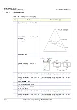
Table 4-21 CFM Noise Floor Test (3S)
Step
Task
Expected Result(s)
1
With the 3S probe in the air, select a Cardiac
preset and activate CFM.
2
Set the CFM ROI to its maximum size.
3
Set the following CFM parameters:
•
Range: 20cm
•
Tissue Priority: 0
4
Set the Active Gain until a few color noise dots
appear in the ROI.
The Active Gain should be between 61and 65.

