©
Home Training Tools Ltd. 2006 Page 6 of 8
Visit us at ww.homesciencetools.com
Ideas for Using Your Microscope
You have a microscope—now what? With
the following directions you can get started right
away making your own microscope slides!
How to Make Simple Microscope Slides
Learn more about using the Kids
microscope by making simple slides using
common items from around the house!
Materials Needed:
-
clear Scotch tape
-
a few granules of salt, sugar, ground
coffee, sand, or any other grainy
material
Making Simple Slides
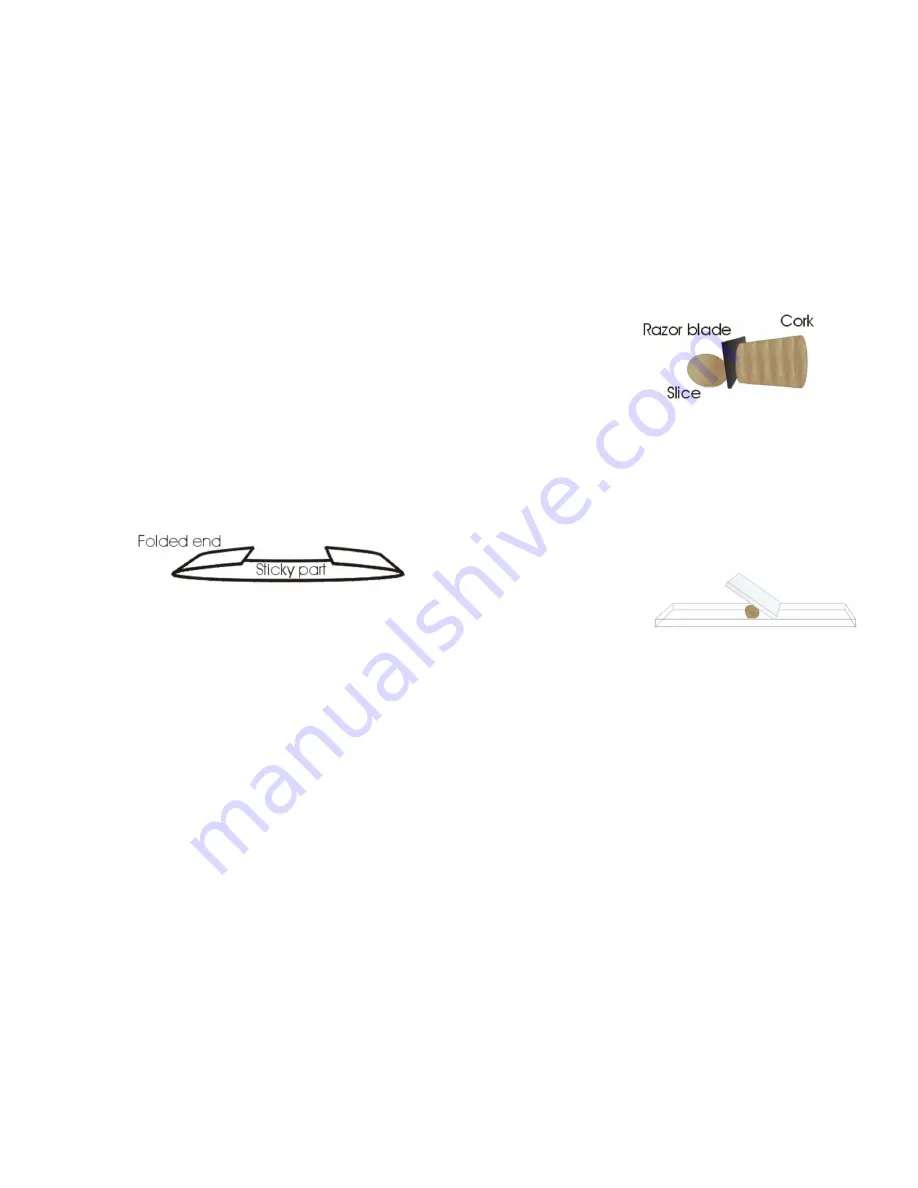
To make a slide, tear a 2½-3” long piece of
Scotch tape and set it sticky side up on the
kitchen table or other work area. Fold over about
½” of the tape on each end to form finger holds
on the sides of the slide. Next, sprinkle a few
grains of salt or sugar in the middle of the sticky
part of the slide.
You can repeat this with the other
substances if you like, just be sure to label each
slide you make with an ink pen or permanent
marker so you will know what’s on the slides!
You can make tape slides with many other
materials as well. Try hair (from pets and family
members), thread and fiber (from carpets or
clothing), or small dead insects such as gnats,
ants, or fruit flies. Label each slide and view
them one at a time with your microscope,
experimenting with different magnification.
How to Make Your Own Prepared Slide
Learn how to make temporary mounts of
specimens and view them with your microscope.
Below are a few ideas for studying different
types of cells found in items that you probably
already have around your house.
Cork Cells
In the late 1600s, a scientist named Robert
Hooke looked through his microscope at a thin
slice of cork. He noticed that the dead wood was
made up of many tiny compartments, and upon
further observation Hooke named these empty
compartments cells. It was later known that the
cells in cork are only empty because the living
matter that once occupied them has died and
left behind tiny pockets of air. You can take a
closer look at the cells, also called lenticels, of a
piece of cork by following these instructions.
Materials Needed:
-
small cork
-
plain glass microscope slide
-
slide coverslip
-
sharp knife or razor blade
-
water
How to make the microscope slide:
Carefully cut a very thin slice of cork using
a razor blade or
sharp knife (the
thinner the slice, the
easier it will be to
view with your
microscope). To
make a wet mount of the cork, put one drop of
water in the center of a plain glass slide – the
water droplet should be larger than the slice of
cork. Gently set the slice of cork on top of the
drop of water (tweezers might be helpful for
this). If you are not able to cut a thin enough
slice of the whole diameter of the cork, a smaller
section will work.
Take one coverslip and hold it at an angle
to the slide so that one
edge of it touches the
water droplet on the
surface of the slide.
Then, being careful not to move the cork
around, lower the cover slip without trapping any
air bubbles beneath it. The water should form a
seal around the cork. Use the corner of a paper
towel to blot up any excess water at the edges
of the coverslip. To keep the slide from drying
out, you can make a seal of petroleum jelly
around the cover slip with a toothpick. Begin
with the lowest-power objective to view your
slide. Then switch to a higher power objective to
see more detail. Use this same wet mount
method for other specimens such as cheek cells
or leaf cells.
Record Your Observations
Our Microscope Observation worksheet (on
the next page) will help you keep track of what
you see and remember what you have learned.
Blanks are provided for recording general
information about each slide (e.g. wet mount
stained with methylene blue). In addition, there
is space to write down your observations and
make sketches of what you see at each
magnification level.


