8/9
9
MICROSCOPE GLOSSARY
'DUNILHOG0LFURVFRS\
Darkfield microscopy is a method of acquiring a slide image. It is specific to the way the light is projected onto a
slide in the microscope equipped with darkfield condenser. The method allows elements in the field of view to be
distinguished that would be invisible or poorly visible using other methods of lighting (such as brightfield or phase
contrast light). This method is commonly used to evaluate slides on which the object and the background are the
same colour. (Such as red blood cells in vital blood or clear bacterial organisms in saline.) In darkfield microscopy,
objects appear in the form of glowing outlines on a black/dark background.
The advantage of darkfield illumination is that details can be seen that are normally not resolved by the
microscope’s objective. While the actual details cannot be seen, the light reflected by them can. A nice analogy here
is that of dust in a room. Normally, in a well-lit room very small dust particles cannot be seen. However, if the lights
are turned out, a beam of light from an acute angle makes these same particles visible. Besides the optical
advantages, darkfield illumination is very beautiful and gives an almost science-fiction-like image.
What is the difference between darkfield and brightfield?
The difference between the two is the way light is projected onto the slide. As a result, elements on the slide not
visible (or poorly visible) in bright light are visible with darkfield illumination.
%ULJKWILHOG
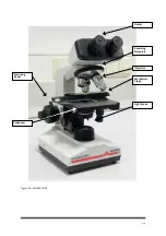
This is the basic microscope configuration.
In brightfield microscopy, the transparent or translucent specimen is either naturally coloured or stained and
appears dark against a bright, white background or field.
3KDVH&RQWUDVW
Phase contrast is a technique for revealing the structural features of microscopic, transparent objects that cannot
otherwise be accomplished with brightfield microscopy. Phase contrast achieves the same effect as staining a
specimen (which can kill a live specimen). In phase-contrast microscopy, the “staining” is in the optics.
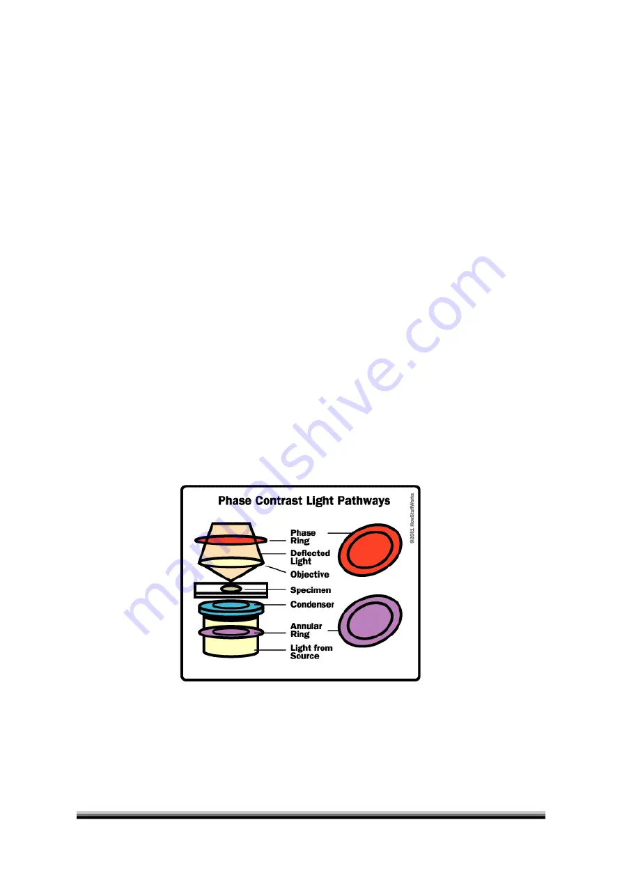
In a phase-contrast microscope, the annular rings in the objective lens and the condenser split the light. The light
that passes through the central part of the light path is recombined with the light that travels around the
periphery of the specimen. The interference produced by these two paths produces images in which the dense
structures appear darker than the background.


