JPK Instruments NanoWizard
®
Handbook Version 2.2a
33
Protect the cantilever
Of course it is important to use clean, fresh cantilevers for high resolution imaging. it is
also important to protect the cantilever once the imaging starts. If the tip is
contaminated with a lot of protein at the beginning, then it will be difficult to get high
resolution images when all the settings are optimized. So start with moderate scan
regions (e.g. 1 - 3 microns), try to avoid too many or too large overview scans, and be
sure to start with the lowest force possible. If the cantilever meets a large particle or
piece of contamination, it may be best to stop the scan and zoom into the already
scanned clean area, or choose another area.
6.2 Imaging hints – intermittent contact mode in liquid
Intermittent contact mode is often chosen for single molecule imaging, because the
molecules may only loosely be stuck to the surface. The lateral forces in contact mode
then tend to sweep the molecules to the side, cleaning the area that is being imaged
and depositing material around the edges.
Cantilevers
Nearly all contact mode cantilevers can also be used for intermittent-contact mode in
liquids, but for high resolution imaging the sensitivity of the cantilever is very important,
and some cantilevers can give much better results. Generally the softest contact mode
cantilevers are too soft for good dynamic mode imaging, because the resonance is very
broad (the quality factor is very low, around 1) and hence the sensitivity is low. The
resonant frequency of very soft cantilevers is also too low for good imaging – for
example, a cantilever with a spring constant around 0.1 N/m often has a resonance of
only 1-5 kHz. The pixel rate must be considered – for instance 512 pixels on trace and
retrace, 1Hz line rate gives 1kHz pixel rate. It is impossible to make a reasonable
amplitude measurement at this rate if the cantilever itself is only oscillating at 1Khz!
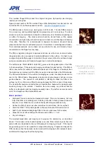
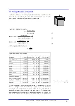
Protein molecules on silicon
Good cantilevers for intermittent contact mode in liquid are usual medium stiffness
(around 0.3 – 0.5 N/m), short (around 100 microns length) and often triangular. Silicon
nitride cantilevers are often available in either normal or oxide-sharpened versions. For
single molecule imaging the oxide-sharpened version can make a significant
improvement to the image quality, since the features are almost exclusively limited by
the tip diameter. A 5 nm tip radius gives a large improvement over a 10-20 nm.
Settings
Use a driving frequency close to the first natural resonance of the cantilever. The
resonant frequency can be conveniently seen by using the thermal noise measurement
tool. It is generally not recommended to measure the sensitivity first – the force
spectroscopy could damage the tip for high resolution imaging. A sensitivity value of
around 50-100 nm/V is typical, and an exact sensitivity is not required, as only the
frequency, not the actual spring constant is interesting here from the thermal noise
measurement. Cantilevers like the ones recommended here have a resonant frequency
around 10 - 13 kHz in liquid.
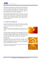
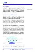
Twisted protein fiber on mica
The oscillation amplitude should generally be low for sensitively imaging small objects.


