JPK Instruments NanoWizard
®
Handbook Version 2.2a
39
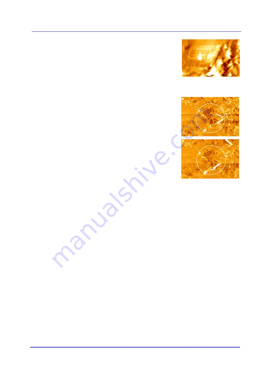
Dull or dirty tips generally lead to features on the surface that have the same shape
and orientation. In this case, what is imaged is actually the shape of the tip, and the
sharper features in the sample act as a probe.
7.3 Contamination
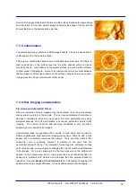
The sample itself may contribute to SPM image artifacts, if there is loose debris or
contamination from the sample surface.
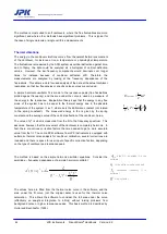
This polymer coated glass slide shows a well-defined sub-structure, but there is
also loose debris on the surface (seen as the white streaks), which is moved
around by the tip. Loose debris on the sample surface is moved by the cantilever
to other areas of the sample. Some of the sample structure is constant between
the two images, but the higher debris on the surface changes from scan to scan.
(Image sizes 25 x 25 µm, intermittent contact mode)
7.4 Other imaging considerations
Tip shape and interaction forces
Since an interaction force is measured by the cantilever, then the probe always
also exerts some force on the sample. This can cause problems of distortion or
damage to the sample, which may move under this force, particularly since many
biological samples are soft and delicate, and require particularly careful AFM
imaging. The choice of probe and scanning conditions is very important,
depending on the sample to be imaged.
Commercial probes are available with a variety of cone angle and tip radii for
different applications; high resolution imaging will require a sharp tip with a high
aspect ratio to accurately reproduce the sample. There are other artifacts that
commonly occur in scanning, however, in addition to the ones from the
geometrical shape of the tip. The interaction forces may also compress or drag
parts of the sample, moving objects or changing the height or width of soft features
in the images. For a given imaging force, the local pressure on the surface will
depend on the contact area, and in some cases the use of ultra sharp tips can
cause more problems with distortion and damage than the potential benefit in
resolution. See also
Section 5.5
Section 5.6
for a discussion of imaging soft
samples with a large height difference, for the specific example of cell imaging.


