8
User
'
s Manual Ophthalmoscope
KaWe EUROLIGHT
®
E25 / E35 LED, 5 apertures
Dear Customer, thank you for choosing a KaWe pro-
duct. Our products are known for their high quality
and long life. This KaWe product meets EC standards
93/42/EWG (standards for medical products).
Please read these instructions thoroughly and
carefully before attempting to use this product
and heed the given instructions for the care of
the product!
Familiarize yourself fully with the operation
of this product before attempting to use it.
Use:
These ophthalmoscopes are only to be used by
authorized professional personnel. They are only to
be used for the purpose of examining the fundus of
the eye.
Purpose:
KaWe ophthalmoscopes are used to exa-
mine the fundus of the eye using direct ophthalmos-
copy. The detection of characteristic changes in the
fundus of the eye enables the diagnosis of numerous
diseases. The conceptual design of the KaWe ophthal-
moscope allows for its use without pupil dilating
medication. In this case, however, the patient’s field of
sight is limited.
Unsuited use/contraindication of the products:
Any use other than that described here is not in accor-
dance with the intended use of the unit. The manufac-
turer is not liable for any resulting damages. The user
alone bears the risk. To power the KaWe EUROLIGHT
®
E25, use exclusively the KaWe MedCenter
®
5000 wall
charging station.
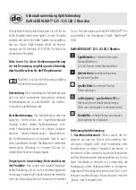
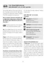
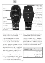
KaWe EUROLIGHT® E25 / E35 LED,
5 apertures
Slit aperture
for the detection
of height fluctuations
(from tumors or papilledema etc.)
Small spot
for normal fundus examinations
Large spot
for normal fundus examinations
Red-free (green)- large spot RF
for detecting minute vein abnormalities,
filters red light waves
from the observation field
Fixing agent
for detecting central
and excentric fixation
Operation of the Ophthalmoscope
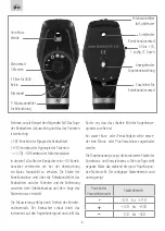
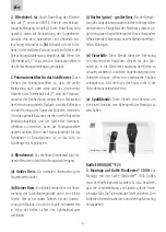
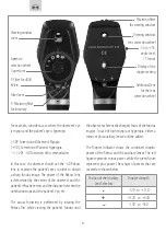
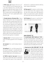
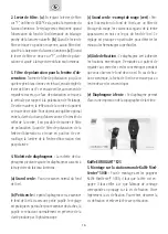
1. Rekoss Lens:
If both the observer and the patient
are completely emmetropic (have perfect vision),
it should be possible to obtain a clear fundus image
without the use of a corrective lens. However, in most
cases, both the observer’s and the patient’s eyes have
refractive errors and in order to obtain a clear fundus
image, it is necessary to correct the combination of
these refractive errors. Hyperopic vision is corrected
with a “plus” lens while myopic vision is corrected with
a “minus” lens.


