background image

3.       Do not disassemble any part of the microscope as this could result in malfunction or mitigated performance.
4.   

When not using the microscope, ensure that the frame is fully cooled before storing the unit in a dry locker or 
covering with a dust cover (provided).

5.   

To clean the Flip Top Condenser, fully loosen the securing thumb screw (1) and remove the condenser then, wipe 
the front lens of the condenser with optical cleaning solution (mixture suggested above) and lens tissue.


The Flip Top Abbe can be re-attached in its seat, by tightening securing thumb screw, and raising condenser 
bracket to desired position. (As shown in picture)


6.   

Be sure to observe you local rules / regulations for product disposal.

7.   

Always cover the Microscope with the provided dust cover when not in use

II)     Optical Cleaning

1.   

The  objectives  have  been  adjusted  for  a  tight  fit  to  prevent  any  damage  during  transportation. To  remove  an 
objective, rotate it counterclockwise while gripping it with a rubber sheet, etc. to avoid any slippage.

2.   

To clean the lens surfaces, remove dust using a soft brush or compressed air (cans available at your local electronic 
store). For removing finger marks or grease, soft cleaning cloth or lens tissue lightly moistened wit cleaning solution 
(85% petroleum ether and 15% isopropanol) should be used. For cleaning the objective optics, use Meathanol. 
Observe sufficient caution in handing Methanol. Place the Objectives and / or eyepieces on a dust free surface (e.g. 
aluminium foil). All other optical components to be cleaned should be as accessible as possible.

3.   

Blow all loose dust particles away with compressed air of mini dust blower.

4.   

Remove all water-soluble dirt with distilled water. If this is unsuccessful repeat using a solution of diluted hand soap 
liquid. Remove any remaining residue with a dry cotton slab.

5.   

To remove oil, use a solution of diluted hand-soap liquid initially. If this does not produce a satisfactory result, repeat 
the cleaning using a solvent (Optical Cleaning Solution 85% petroleum ether and 15% isopropanol).

6.   

Grease must always be removed using a solvent.

7.   

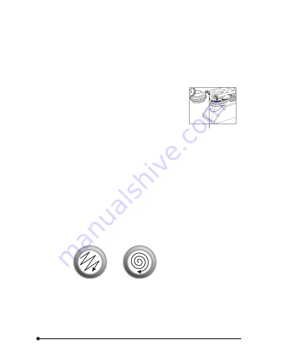
Cleaning is achieved by using a spiral motion from the center to the rim. Never wipe using zig-zag movements as 
this will only spread the dirt. With larger optical surfaces (e.g. tube lenses) the spiral motion starts initially at the rim 
before moving to the middle and is then followed by a center to rim cleaning motion. Normally several spiral wipes 
are recommended.


We recommend pure, volatile petroleum ether or Optical Cleaning Solution as explained in point 3 above.


            Wipe using a spiral movement       Donot use a zig-zag motion

III)     Cleaning of Painted Surfaces

Avoid the use of any organic solvent (e.g. thinner, xylene, ether, alcohol etc.) for cleaning of painted surfaces of the 
instrument. Painted surfaces can be cleaned with a very lightly moistened micro fiber cloth. Loose dust and other dirt 
particles can be removed using a soft bristle brush used exclusively for this purpose.

3

1

Summary of Contents for Lx POL

Page 1: ...re proper use of this instrument as well as to avoid injury while operating instrument understanding this manual completely before use is highly recommended User Manual Lx POL Research Microscopy R R...

Page 2: ......

Page 3: ...YOUR MICROSCOPE 8 4 Lx POL TRINOCULAR 7 3 Lx POL BINOCULAR 6 7 OPTIONAL ACCESSORIES INSTALLATION 10 11 8 DETAILED INTRODUCTION OF ASSEMBLIES 12 14 a Flip Top Abbe Condenser b Round Stage c Centerable...

Page 4: ...t 11 SUMMARY OF POLARISED OBSERVATION PROCEDURE 20 12 SUMMARY OF POLARISED OBSERVATION PROCEDURE 21 24 a Placing the specimen on stage b Adjusting the focus c Adjusting the Interpupillary Distance d A...

Page 5: ...bjectives The round ball bearing Rotatable Stage has smooth 360 degree travel and has 1 degree graduations on the scale which provides accurate location of the specimen High power illumination is deli...

Page 6: ...Lx POL if the microscope is used in a manner not specified by this manual the safety of the user may not be warranted In addition the equipment may also suffer damage Always use the equipments as out...

Page 7: ...ng Methanol Place the Objectives and or eyepieces on a dust free surface e g aluminium foil All other optical components to be cleaned should be as accessible as possible 3 Blow all loose dust particl...

Page 8: ...oscope on a sturdy level table or bench and aovid any restriction of air vents in the base of the unit Do not place microscope on a flexible surface as this could result in blocking the air vents and...

Page 9: ...bulb reduce its life If contamination occurs wipe bulb surface with a cloth slightly moistened with alchohol Applicable Bulb 6V20W Halogen Bulb P N CX 013 Fig 1 Caution For Fuse replacement Set the m...

Page 10: ...der Vernier Scale Flip Top Abbe Condenser Polarizer Koehler mount Coarse and fine focus knob Polarizer locking screw Intensity regulator Bertrand lens focusing knob Bertrand lens on off Allen keys for...

Page 11: ...king screw Circular stage with vernier Port of wave plate Analyzer on off Parking for additional wave plates 2 Allen keys for objective centering Trinocular viewing tube 30 inclined Bertrand lens focu...

Page 12: ...8 UNPACKING YOUR MICROSCOPE 5 Microscope Arm Observation head Eyepieces Power Cord...

Page 13: ...Paired Eyepieces 6V 20W Halogen bulb R Binocular viewing tube 30 inclined Eyepieces Intermediate Analyzer Kit Bertrand Lens centering screw Centerable Revolving Nosepiece Polarizing Objectives Specime...

Page 14: ...ories OPTIONAL ACCESSORIES 7 Binocular head Flip Top Abbe Condenser Blue filter RP 2 5x RP 10x RP 20x RP 40x SL RP 100x SL Oil Trinocular head WF 10x WF 16x WF 20x Halogen Bulb iVu 5100 iVu 7000 R R R...

Page 15: ...ar observation head Fig 5 2 Mount iVu Camera Module System Fig 3 on video adapter 1 iVu Camera Module System 10X eyepieces are provided To replace 1 Pull out the 10x eyepieces out from the observation...

Page 16: ...scope s optical axis This makes the center of rotation coincide with the center of field of view Rotatibility allows the user to observe the specimen in the diagnol position the brightest position of...

Page 17: ...another Lx POL Nose Piece is fully equipped with centering mechanism for each objective to make sure that each object is centered to the stage and microscopic optical axis so that the specimen remains...

Page 18: ...rience Fig 13 14 1 Bertrand Lens Focusing Knob 2 Bertrand Lens Operating Lever 3 Analyzer Operating Lever 4 Gypsum wave Plate 5 Allen wrench for Objective centering 6 Allen screw for Bertrand Lens Cen...

Page 19: ...Locking Screw 1 See figure 15 3 Remove the Dust Cover Cap from Observation Head Dovetail and mount the Observation Head by engaging the dovetail provided at the bottom of the head into the dovetail c...

Page 20: ...b in the opposite direction decreases brightness The intensity bar next to the knob indicates the direction of intensity level Note Never use an adapter between the power cord and the power source it...

Page 21: ...diaphragm so that its closed iris leaves image is present within the field of view Use the condenser focusing knob to bring the image into sharp focus Operate the condenser centering screws simultane...

Page 22: ...ntil the prominent point of the specimen is furthest away from the center of the Eye Piece crossline it may even be outside the field of view Fig 24 b iii Adjust the image with the centering screws ov...

Page 23: ...n 1 Bring an objective of your choice between 20x to 100x in the light path Bring the Pol specimen into focus 2 Keep the abbe condenser in its lowest position 3 Swing inTop lens of FlipTopAbbe Condens...

Page 24: ...o Eye Piece cross hair scale Adjust the observation tube and Adjust the interpupillary distance Adjust the dioptric setting Engage Polarizer Analyzer filters in the light path and use Bertrand Lens an...

Page 25: ...g position The degree of position can be noted Vernier Scales These scales allow for easy identification of the specimen s position coordinates making it easy to return to a particular region of inter...

Page 26: ...tereo vision This greatly helps to reduce fatigue and discomfort during observation While looking through the eyepieces move both eyepieces laterally until the left and right fields of view coincide c...

Page 27: ...nto focus 3 While looking through the left eyepiece with your left eye rotate only diopter adjustment ring on the eyepiece until specimen is at its best possible focus The condenser is most often used...

Page 28: ...to focus Since air bubbles in the oil will affect the image quality make sure that the oil Is free of bubbles To remove bubbles rotate the revolving nosepiece slightly to agitate the oil 4 The condens...

Page 29: ...th an Use immersion oil as suggested immersion objective Bubbles are present in immersion oil Remove the bubbles by agitation The specified immersion oil is not used Use the immersion oil supplied by...

Page 30: ...Check and replace with live fuse 10 Bulb blows easily The specified bulb is not used Replace with the specified bulb 11 Field remain dark even Bulb is on In the Exintetion Position Remove Analyzer fr...

Page 31: ...ty 360 degree Specimen holder Spring Loaded stage clips 6 Condenser Type Flip Top Abbe condenser daylight filter detachable N A 1 25 Aperture iris diaphragm Built in 7 Dimensions Weight 405mm L x 210m...

Page 32: ...09 LABOMED and Lx POL are registered trademarks of Labo America Inc With a policy of continuous development Labo America Inc reserves the right to change design and specifications without prior notice...

Reviews: