54
4.9 Centring the aperture and field diaphragm
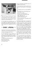
Centring the aperture diaphragm
Turn a low to medium objective magnification
10x/20x into the light path and focus a strongly
reflecting specimen (e.g. surface mirror) via the
BF reflector with the coarse and fine drive.
Remove an eyepiece from one of the two eye-
piece tubes and look into the empty tube.
Regulate the light intensity so that the rear objec-
tive pupil (rear lens surface of the objective) can
be clearly seen.
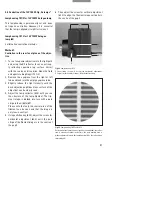
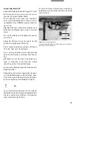
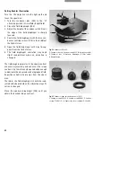
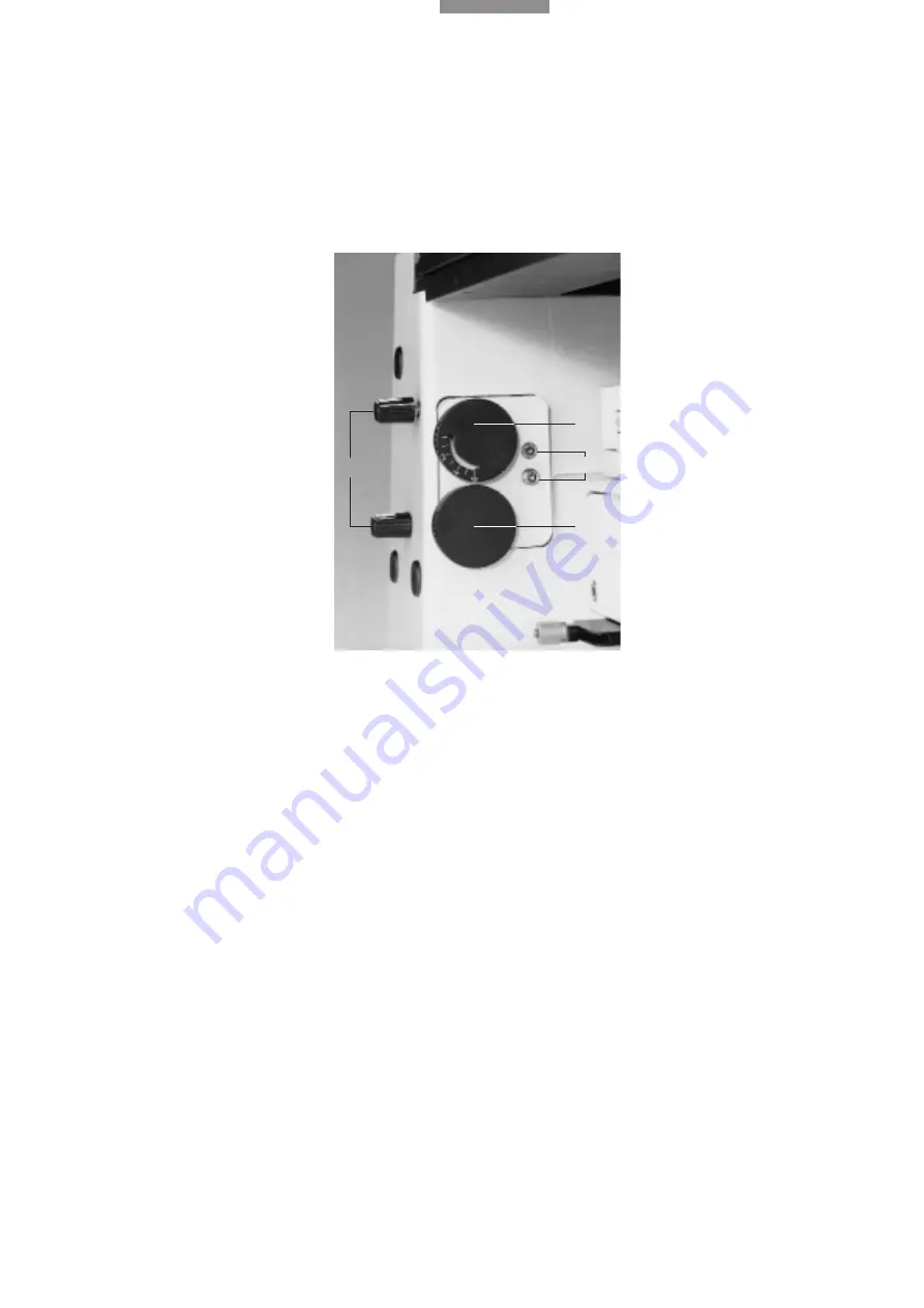
Using the adjustment button (72.1), open the
aperture diaphragm nearly to the edge of the
pupil.
Centre the aperture diaphragm to the edge of the
pupil with the centring screws (72.2).
The aperture diaphragm influences the resolu-
tion, contrast and field depth of the microscope
image. Image quality greatly depends on how
carefully it is set. It may not be used for regulating
the image intensity.
Centring the field diaphragm
Turn a low to medium objective magnification
10 x/20 x into the light path and focus a strongly
reflecting specimen (e.g. surface mirror) via the
BF reflector with the coarse and fine drive.
Open the field diaphragm almost as far as the
edge of the field of view.
Using the centring buttons (72.4), centre the field
diaphragm to the edge of the field of view.
The field diaphragm is imaged on the surface of
the specimen, framing the illuminated field.
Normally, the field diaphragm is opened until it
just disappears out of the field of view.
When imaging reduced picture diagonals such
as in photomicrography or TV microscopy, the
field diaphragm can be narrowed to frame the
picture format, enhancing the image contrast.
The aperture diameter of the field diaphragm
remains the same for all objective magnifica-
tions.
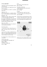
Fig. 72
Aperture and field diaphragm
1
Aperture diaphragm adjustment,
2
Aperture diaphragm centr-
ing screws,
3
Field diaphragm adjustment,
4
Field diaphragm
centring screws
1
3
2
4


