Esophageal Stent Technology System™
DEVICE DESCRIPTION
The MERIT ENDOTEK™ ALI
MAXX-ES™
Esophageal Stent
System is comprised of two components: the radiopaque
self-expanding nitinol stent and the delivery catheter.
The stent is completely covered with a biocompatible
polyurethane membrane and a silicone coated inner-
lumen. The stent expansion results from the physicial
properties of the metal and the proprietary geometry.
The stent is designed with a some what larger diameter
at the distal and proximal ends to reduce the possibility
of migration. The overall stent geometry is designed to
minimize foreshortening upon expansion, thus facilitating
improvement in deployment accuracy. The proximal end
of the stent is threaded with a suture intended for use
in proximal repositioning of the stent. (See description
under REPOSITIONING OF THE ESOPHAGEAL STENT).
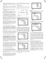
The stent is deployed with a dedicated delivery system.
The delivery system consists of two coaxial sheaths
attached to a deployment handle. The handle permits
one-handed positioning and deployment via a trigger
mechanism. The exterior sheath serves to constrain the
stent until the sheath is retracted during deployment.
Once deployment is initiated, the stent
can not
be
reconstrained. An indicator on the handle mechanism
provides the operator with tactile feedback when the
stent has been deployed to 50% of its length.
This is
the last point at which the operator can reposition
the stent proximally by pulling the entire delivery
catheter proximally.
A radiopaque tip and marker on
the inner shaft aid the operator in determining stent
position in relation to the deployment threshold, where
repositioning or en bloc withdrawal is no longer possible.
The inner tube of the coaxial sheath catheter contains
a central lumen that will accommodate a 0.035” guide
wire. This feature is designed to allow safe guidance of
the delivery system to the intended implant site while
minimizing the risk of esophageal injury from the delivery
system tip.
The complete Instructions for use should be reviewed
before using this system.
INDICATIONS FOR USE
The MERIT ENDOTEK™ ALI
MAXX-ES™
Esophageal Stent is
intended for maintaining esophageal luminal patency in
esophageal strictures caused by intrinsic and/or extrinsic
malignant tumors and for occlusion of esophageal fistulae.
The stent is also indicated for stenting refratory benign
esophageal strictures for up to 6 months.
CONTRAINDICATIONS
The MERIT ENDOTEK™ ALI
MAXX-ES™
Esophageal Stent is
contraindicated in:
1. Patients with significantly abnormal coagulopathy.
2. Patients with necrotic, chronically bleeding or
polypoid lesions.
3. Strictures that cannot be safely dilated to allow
passage of the delivery system.
4. Esophageal fistulae or perforation that prevent
secure stent placement.
5. Situations that require positioning the proximal end
of the stent within 20mm of the upper esophageal
sphincter.
6. Patients in whom endoscopic procedures cannot be
safely performed.
7. Any use other than those specifically outlined under
Indications for Use.
POTENTIAL COMPLICATIONS
Complications have been reported in the literature for
esophageal stent placement with both silicone stents
and expandable metal stents. These include, but are not
necessarily limited to:
PROCEDURAL COMPLICATIONS:
• Bleeding
• Esophageal perforation
• Pain
• Aspiration
POST-STENT PLACEMENT COMPLICATIONS:
• Stent migration
• Perforation
• Bleeding
• Pain/foreign body sensation
• Occlusion due to lesion growth
• Obstruction related to food volume
• Infection
• Reflux
• Esophagitis
• Esophageal ulceration
• Edema
• Fever
• Fistula formation outside of normal
disease progression
• Death with cause outside of normal
disease progression
ADDITIONAL CAUTIONS AND WARNINGS
1. The MERIT ENDOTEK™ ALI
MAXX-ES™
Esophageal
Stent System should be used with caution after
careful consideration of the following:
• Stent placement across the gastro-esophageal
junction may increase migration risk and reflux.
• Stent placement may further compromise patients
with significant cardiac or pulmonary conditions.
• Laser ablation of lesions with a stent in place could
cause patient injury.
• Placement of a second stent within the lumen of
another stent could significantly compromise the
patency of the lumen.
• Placement of a stent in a very proximal location
could cause discomfort or patient foreign body
sensation.
• Stents placed to treat strictures where the
proximal margins are located within 45mm of the
upper esophageal sphincter may not fully expand,
compromising the patency of the lumen.
2. If the stent is damaged or does not fully expand
during implantation, remove the stent following the
directions for use.
3. Do not cut the stent or delivery catheter. The device
should only be placed and deployed using the
supplied catheter system.
4. Do not reposition the stent by grasping the
polyurethane covering. Always grasp the suture
knot or a metal strut to reposition the stent and do
not twist or rotate the stent or metal strut unless the
stent is being removed.
INSTRUCTIONS FOR USE
Required Equipment:
• Endoscope
• 0.035” (0.89mm) stiff bodied, soft tipped guide wire,
180cm length minimum
• ALI
MAXX-ES™
Esophageal Stent of appropriate
length and diameter
• Fluoroscopic imaging should be used to facilitate
esophageal dilation if required prior to stent
placement. Fluoroscopic imaging may also be used
in addition to or in place of endoscopy to aid in
accurate stent placement.
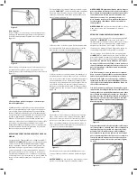
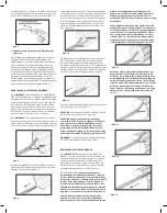
1. Locate Stenosis and Pre-Dilate as Necessary.
Pass an endoscope into the esophagus and beyond
the esophageal stricture. If necessary, dilate the
stricture until an endoscope can be passed.
WARNING:
Do not attempt placement of the MERIT
ENDOTEK™ ALI
MAXX-ES™
Esophageal Stent in patients
with stenoses that cannot be dilated sufficiently to allow
passage of an endoscope.
2. Estimate the Stenosis Length and Luminal
Diameter.
This estimation may be performed by visual inspection
via endoscopy or via fluoroscopy. To determine the
stenosis length, measure the distance from the distal
border of the narrowing to the proximal border while
pulling back on the endoscope. A suitable length estimate
may be obtained with a combination of endoscopy,
fluoroscopy, and a radiopaque marker of known length
that is adhered to the patient’s chest. To determine the
lumen diameter, estimate the diameter of the normal-
appearing esophageal lumen proximal to the stenosis. An
open biopsy forceps may be used for a reference guide.
Alternatively, the stenosis length and luminal diameter
may be measured by reviewing a recent CT Scan of the
narrowed esophageal lumen.
3. Identify Landmarks to Aid in Placement.
Endoscopically and or fluoroscopically examine the lumen
both proximal and distal to the stenosis. The stricture
should be dilated to allow passage of an endoscope,
or approximately 9mm (27F) minimum. Radiopaque
markers may be placed on the patient’s chest to assist in
identifying the margins of the stenotic area.
4. Select the Appropriate Covered Stent Size.
The physician should select a stent diameter following
the complete endoscopic and fluoroscopic examination.
To minimize the potential of stent migration, dilate the
stricture ONLY if passage of the endoscope or the delivery
system through the stricture lumen is not possible.
Choose a stent long enough to completely bridge the
target stenosis with a 25mm margin both proximally
and distally. Because the MERIT ENDOTEK ALI
MAXX-ES™
Esophageal stent will not significantly foreshorten when
deployed it is not necessary to account for shortening.
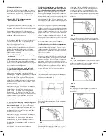
5. Introduce the Guide Wire.
Place a 0.035” (0.89mm), stiff-bodied, soft-tipped
guidewire through the endoscope and beyond the
stenosis. The endoscope should be removed at this time
while maintaining the position of the guide wire.
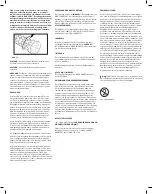
6. Inspect and Prepare the ALIMAXX-ES™
Esophageal Stent System.
This product is supplied non-sterile. Before opening the
package, inspect the package for damage. Do not use if
the package has been opened or damaged.
Carefully remove the device from the plastic packaging
backing card. Visually inspect the Esophageal Stent and
the delivery catheter for any sign of damage. Do not use if
there are any visible signs of damage.
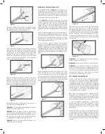
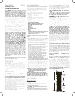
Overall Stent
Length
Stent
Midbody
Distal
Flare
Proximal
Flare
Suture
Knot

