10 Physiological Unit Signal
Operator’s Manual
10 - 3
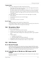
Triggering Mode
There are three triggering modes available: Single, Dual, and Timer.
•
Single Trigger:
When an R waveform is detected, an image will be triggered after delay time
T1. The time of T1 can be edited in single mode.
•
Dual trigger: when an R waveform is detected, two images in two windows will be triggered
respectively after delay time T1 and T2. The time of T1 and T2 can be edited in dual mode.
•
Timer Trigger: an image will be triggered after a time interval.The time interval can be edited
in triggering status.
The image triggering operation is described as follows (Take single trigger as an example):
1.
Select exam mode.
2.
Tap [Trig Mode] on the touch screen to enable the trigger.
3.
Select [Single].
4.
Set the delay time (or use the T1 by default).
Real & Trigger
Tap [Real & Trigger] to enable or disable the real trigger function.
After the [Real & Trigger] is enabled, two images are displayed respectively in two windows. One
is triggered by ECG, and the other is non-triggered real time image.
10.2 Respiratory Wave
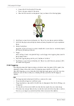
Perform the following procedure:
1.
Connect the ECG lead and position the ECG electrodes.
2.
Tap [Physio] or press the user-defined key for “Physio” to enter Physio screen.
3.
Switch the imaging modes and display formats, adjusting the parameters to get an optimized
image.
4.
Parameter adjusting:
a.
Tap [RESP] on the touch screen.
b.
Adjust [Speed], [RESP Gain], [Position] and [Invert].
5.
Exit Respiratory display mode, and remove ECG electrodes from the patient.
6.
Tap [Physio] or press the user-defined key for “Physio” to exit physio mode.
10.3 ECG Review
10.3.1 Review Principle
When an image is frozen, the ECG waveform where the image is triggered will be frozen at the
same time. In the Dual triggering mode, the two window images are frozen at the same time.When
images are reviewed with the ECG electrodes connected, the ECG trace is the reference for time.
After the images are frozen, all real time images are in the status of linked review.
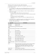
10.3.2 Linked Review of Waveforms, M/D Images and 2D
Images
If the physio unit signal, time curve and 2D image are frozen at the same time, the replay of them is
displayed at the same time.
Summary of Contents for Anesus ME7T
Page 2: ......
Page 58: ...This page intentionally left blank ...
Page 154: ...This page intentionally left blank ...
Page 164: ...This page intentionally left blank ...
Page 182: ...This page intentionally left blank ...
Page 190: ...This page intentionally left blank ...
Page 208: ...This page intentionally left blank ...
Page 254: ...This page intentionally left blank ...
Page 264: ...This page intentionally left blank ...
Page 280: ...This page intentionally left blank ...
Page 311: ......
Page 312: ...P N 046 018839 00 5 0 ...

