Instrument Performance Factors
30
5000499 D
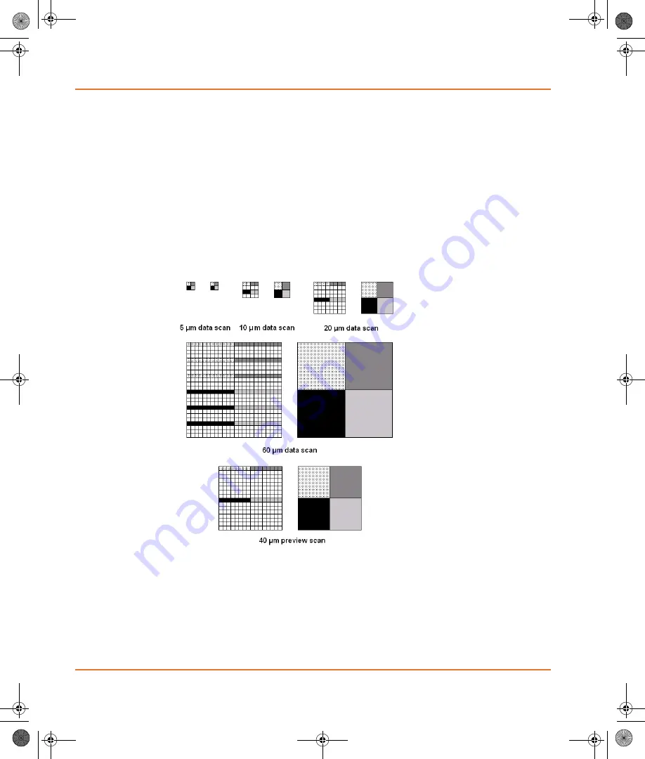
For a preview scan, the same 5
μ
m spots are scanned. However the
Y-direction step size is increased to 40
μ
m, and each 40
μ
m preview
scan pixel is an average of eight contiguous 5
μ
m spots in the
X-direction. This speeds image acquisition for balancing the PMT gains
while minimizing photobleaching.
.
is a representation of how the 4100A Microarray
Scanner samples 5
μ
m spots and averages them to calculate the values
of the image pixels for the various scan modes and pixel sizes. On the
left side of each image pair is the raw data scan from the scanner. Each
square represents a 5
μ
m by 5
μ
m spot as scanned by the 5
μ
m laser
beam. On the right are the resultant image pixels at various
resolutions. The shading indicates the spots that are scanned as well as
the corresponding pixels in the final image.
.
Figure B-1
Data scans
GenePix_4100A.book Page 30 Friday, October 22, 2010 3:21 PM


