10
IMAGING SCENARIOS
WARNING
A patient safety strap must be used during all procedures.
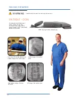
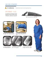
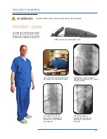
PATIENT - LIZ
Liz in the SPS II while obtaining a
lateral image of the upper thoracic
spine.
Liz in the SPS II while obtaining an
AP image of the upper thoracic spine.
Lateral image primarily through
the C7—T2 segments.
AP image visualizing the
T1-2 interlaminar space.
Contralateral oblique showing
the upper thoracic facet joints.
Components used: Radiolucent Frame,
Crescent Face Pad, Contoured Torso
Support Pad, Contoured Torso Wedge,
8” Semi-Round Bolster (not pictured)
SPS II set up for Liz shown here


