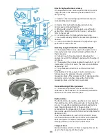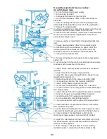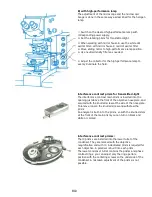
B1
Transmitted-light brightfield microscopy
A) with halogen lamp:
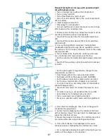
When doing transmitted-light brightfield microscopy with the
halogen lamp, the controls of the Univar are set at the red-
dot positions.
⚪ Turn on halogen lamp with control transformer
⚫ Turn on relay (red dot)
⚫ Switch off Bertrand lens (pull out pin)
⚫ Turn off neutral density filters in the body (black dot not
visible)
⚫ Deactivate blocking-filter slide or rotatable analyzer (red
dot)
⚫ Switch off the compensator
⚫ Pull out interference-contrast main prism until it stops
(loosen the clamping screw on the left side first)
⚫ Place into position the 10x objective
⚫ Turn condenser turret to brightfield wide-field
⚫ Swing out the transmitted-light polarizer.
⚫ Set condenser fine-feed in the central position and move
condenser up to the stop with the coarse feed
⚫ Turn lower condenser turret to the empty opening (red
dot)
⚫ Switch off excitation filter for transmitted-light (red dot)
⚫ Switch off neutral density filters for transmitted-light (red
dot)
⚫ Switch off the auxiliary optics for wide-field condenser (red
dot)
⚫ Adjust collector for transmitted-light halogen lamp set (red
dot)
⚫ Adjust the photo system magnification changer to low
magnification (L, red dot)
⚫ Set the beam path to the camera (red dot, CAM)
⚫ Set the beam to 20% to the tube (red dot, CAM/PRO)
⚫ Switch the phase ring knob to empty (red dot)
⚫ Set the magnification changer to 1x (red dot)
⚫ On mirror housing 2, set the rotary prism on transmitted-
light with halogen lamp
⚫ Switch off color filters for contrast fluorescence (red dot)
⚫ On mirror housing 2 set excitation filter on red dot
⚫ Set sliding mirror for halogen lamp (red dot)
⚫ Turn on automatic zoom lighting (red dot)
⚫ Focus on a specimen with coarse and fine adjustment
⚫ Slightly close field iris diaphragm and focus on the image
with condenser fine adjustment in the specimen plane.
⚫ Center field iris diaphragm with centering screws on the
condenser turret. Then open field iris diaphragm slightly
larger than field of view
⚫ Close the condenser iris diaphragm so that the
microscopic image appears clear and high contrast. This is
usually the case when the oculars' back focal plane has
about 2/3 of their diameters brightly illuminated (check with
Bertrand lens)
⚪ When changing objectives, check the openings of the field
and the condenser iris diaphragms and correct if necessary.
Summary of Contents for Univar
Page 1: ...Reichert Univar Manual...
Page 2: ......
Page 48: ...D6 blank no content...
Page 58: ...E2 blank no content...


