B3
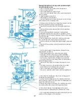
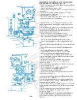
Transmitted-light darkfield microscopy
A) with halogen lamp:
⚪ Switch to the halogen lamp with control transformer
⚪ Relay switch: red dot
⚪ Turn off the Bertrand lens (pull out pin)
⚪ Switch out the eyepiece neutral density filters (black dot not
visible)
⚫ With the immersion darkfield condenser, use the 40x objective
⚫ With the dry darkfield condenser, use the 25x objective
⚪ Switch off exciter filter for transmitted-light (red dot)
⚪ Switch off neutral density filters for transmitted-light (red dot)
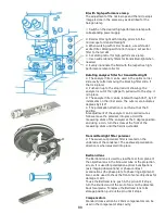
⚪ Use darkfield condenser: apply immersion oil to the immersion
darkfield condenser
⚫ When working with the dry darkfield condenser and the Apo 25x
and Plan 40x objectives, the aperture insert is inserted into the
condenser.
⚪ Swing out the transmitted-light polarizer
⚫ Set condenser fine drive in the central position and move
condenser, with the coarse drive, up to the stop. With the
immersion darkfield condenser, use oil between condenser and the
specimen slide
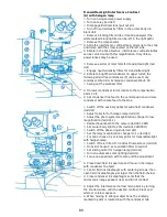
⚪ Turn to an empty opening of the lower condenser turret (red dot)
⚪ Switch off the auxiliary optics (if available) for wide field
condenser (red dot)
⚪ Adjust the collector for transmitted-light halogen lamp (red dot)
⚪ Set the magnification lever in the photo system to low
magnification (red dot, L)
⚪ Redirect the beam path to the camera (red dot, CAM)
⚪ Set eyepiece light to 20% (red dot, CAM/ pro)
⚪ Set the phase ring knob to empty (red dot)
⚪ Set magnification changer to 1x (red dot)
⚪ Disengage blocking-filter slide or rotatable analyzer (red dot)
⚪ When using the mirror house 2, set the rotary prism on
transmitted-light with halogen lamp
⚪ Disengage the compensator
⚪ Interference contrast main prism: loosen the clamping screw on
the left side and pull out to the stop (red dot)
⚪ Switch off color filters for contrast fluorescence (red dot)
⚪ On mirror house 2, set excitation filter to red dot
⚪ Switch sliding mirror to halogen lamp (red dot)
⚫ Switch off the automatic zoom lighting (lever to the side)
⚫ Set manual zoom lighting to DF
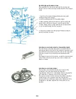
⚫ Fully open condenser iris diaphragm. Focus on the specimen
⚫ Slightly close field iris aperture and focus on the diaphragm
image with the condenser fine adjustment. Frame the field iris
diaphragm in the middle of the field using the centering screws.
Then open the field iris diaphragm just past the field of view
⚪ When moving a different objective into position, the iris of the
objective can be closed to avoid glare by turning the the knurled
ring. This objective iris can be opened again for brightfield work.
Summary of Contents for Univar
Page 1: ...Reichert Univar Manual...
Page 2: ......
Page 48: ...D6 blank no content...
Page 58: ...E2 blank no content...


