C11
Mixed illumination: epifluore transmitted-light
interference contrast
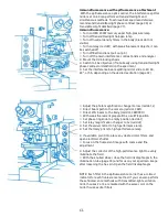
A) Transmitted-light interference contrast with halogen lamp
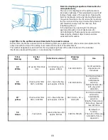
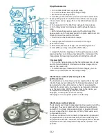
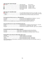
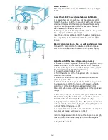
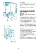
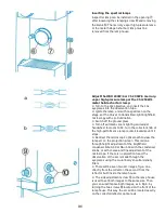
⚪ Turn on halogen lamp
⚪ Turn on relay optics (red dot)
⚪ Turn off Bertrand lens (pull out pin)
⚪ Turn off neutral density filters in the ocular body (black dot not
visible)
⚪ Rotate main prism for transmitted-light
⚫ Set interference beamsplitter to 40
⚫ Turn to 10x objective (with this objective magnification,
interference contrast studies can be performed. Only stress-free (IK
or P) objectives may be used
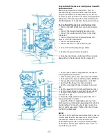
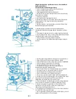
⚪ Switch off exciter or color filters for transmitted-light (red dot)
⚪ Switch on neutral density filters for transmitted-light
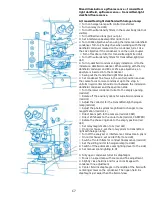
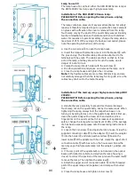
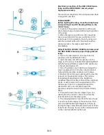
⚫ Rotate upper condenser turret to brightfield condenser. The
following strain-free condensers (P) can be used: dry condenser,
NA=0.90, and immersion, NA=1.30
⚫ Turn lower condenser turret to the compensation prism 10 IC
⚪ Swing off the transmitted-light filter polarizer
⚫ Set condenser fine drive in the central position and raise
condenser with coarse feed up to the stop
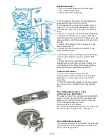
⚪ Switch off the auxiliary optics for wide field condenser (red dot)
⚪ Set collector for transmitted-light halogen lamp (red dot)
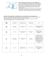
⚪ Adjust the relay magnification to low (red dot, L)
⚪ Direct beam path to the camera (red dot,CAM)
⚪ Set ocular light to 20% (red dot, CAM/PRO)
⚪ Switch off the phase ring knob (red dot)
⚪ Set magnification changer to 1x (red dot)
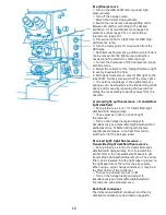
⚪ With mirror house 2, set the rotary prism to transmitted-light with
halogen lamp
⚪ Switch off the color filters for contrast fluorescence (red dot)
⚪ With mirror house 2, set exciter filter off (red dot)
⚪ Set sliding mirror for halogen lamp (red dot)
⚪ Turn on automatic zoom lighting (red dot)
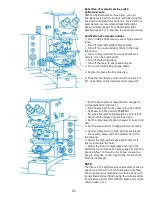
⚪ Focus on a specimen with coarse and fine adjustment
⚫ Slightly close field iris and focus the iris image in the specimen
plane with condenser fine adjustment
⚫ Center the field iris diaphragm in the middle of the field of view
with centering screws. Then open field iris diaphragm just wider than
the field of view
⚫ Close condenser iris diaphragm just until the microscopic image
appears clear and full of contrast, but without diffraction halos
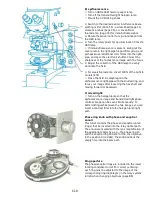
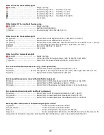
⚪ Adjust the main interference contrast prism by turning the
knurled screw to the desired contrast (black and white or color)
⚪ When switching to a stronger objective, use the appropriate IC
prism in the lower condenser turret
Summary of Contents for Univar
Page 1: ...Reichert Univar Manual...
Page 2: ......
Page 48: ...D6 blank no content...
Page 58: ...E2 blank no content...

