OPERATION
Carl Zeiss
Lighting and contrasting method in transmitted light
Axio Lab.A1
80 430037-7144-001
04/2013
4.1.4.2
Determination of gout and pseudogout
x
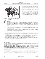
Set the microscope as in the transmitted light brightfield according to KÖHLER (see Section 4.1.1 (3)).
x
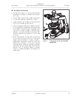
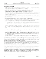
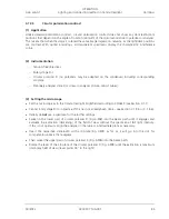
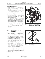
Then swivel the polarizer, rigidly fixed to the lambda plate (445226-0000-000) (Fig. 4-5/
3
) into the
beam path.
x
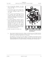
Insert the analyzer slider (Fig. 4-5/
2
) into the slit for compensators.
x
The field of view will appear dark due to the crossed polarizers.
x
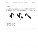
Swivel the rotatable lambda plate into the beam path and set the metal adjusting lever of the lambda
plate to 45°.
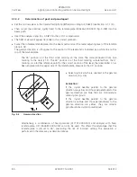
The gamma direction is orthogonal to the position of the level and is indicated by a white line on the
top of the lambda plate.
The 45° position is at the third white marking on the scale. The scale graduation from one
marking to the next is 15°. The 45° position is at the third marking, calculated from the 0°
marking. As a further reference point for the correct position of the lever, the Greek letter
O
has
been engraved on the upper side of the lambda plate, likewise at the 45° position.
x
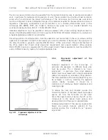
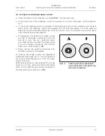
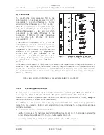
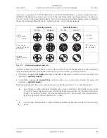
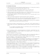
Select crystals which are oriented in the gamma
direction (Fig. 4-6).
Evaluation:
If the crystal needles parallel to the gamma
direction are yellow and those perpendicular to the
gamma direction are blue, they are monosodium
urate crystals (gout).
If the crystal needles parallel to the gamma
direction are blue and those perpendicular to the
gamma direction are yellow, they are calcium
pyrophosphate crystals (pseudogout).
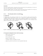
Alternatively, a combination of fixed polarizer (427701-0000-000) and analyzer with fixed
lambda plate, 45° (453681-0000-000) can also be used. This offers the advantage that the
lambda plate is pre-set to 45°, precluding the risk of incorrect setting. The evaluation is
performed in the same way as described above.
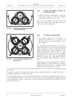
Fig. 4-6
Gamma direction

