6 Detection mode (calculus detection)
Sirona Dental Systems GmbH
Operating Instructions PerioScan
61 29 626 D 3496
20
D 3496.201.02.05.02
*
Root caries is diagnosed through visual inspection (supragin-
gival root caries) or tactile probing (subgingival root caries).
Root caries on proximal surfaces can also be verified with bite
wing exposures. In the visual inspection, the color and contour
of the lesion are evaluated. The main topological difference
between root caries and calculus is that calculus is convex in
shape, while root caries usually exhibits a concave form. This
fact is taken into account when performing tactile probing.
**
A corresponding study was recently published by Meissner et
al (Meissner G, Oehme B, Strackeljan J, Kocher T: "Clinical
subgingival calculus detection with a smart ultrasonic device: a
pilot study" J. Clin Periodontol (35) 2008, p. 126 - 132). Subgin-
gival buccal surfaces of 63 arbitrarily selected periodontally
compromised teeth were scanned intraorally, while the suprag-
ingival positions of the insert, along with the corresponding sig-
nals of the detection system, were saved as separate files. After
extraction, the surface detection results were evaluated by
repositioning the inserts’ position on the tooth in vitro and com-
paring the detection results with visual findings using magnify-
ing glasses. Calculus and cementum were discriminated with a
sensitivity of 91 %, (78.78 % - 96.29 % Clopper-Pearson 95 %
confidence interval) and a specificity of 82 %
(74.81 % - 87.01 % confidence interval).
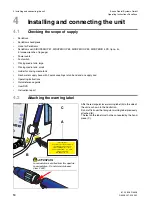
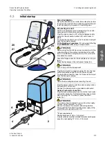
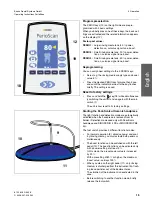
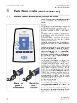
Due to software modifications PerioScan will show you the fol-
lowing results: If PerioScan lightens "green", there is with a
probability of 92 % healthy root surface (confidence interval
90 - 93 %)
***
. If PerioScan lightens "blue", there is with 89 %
calculus (confidence interval 80 - 95 %)
***
.
***
A probability of occurrence of calculus of approx. 20 %
underlies this statement, see Kocher, T., Subgingival polishing
with a teflon-coated sonic scaler insert in comparison to con-
ventional instruments as assessed on extracted teeth, J Clin
Periodont, 27, 243, 2000.
6.2
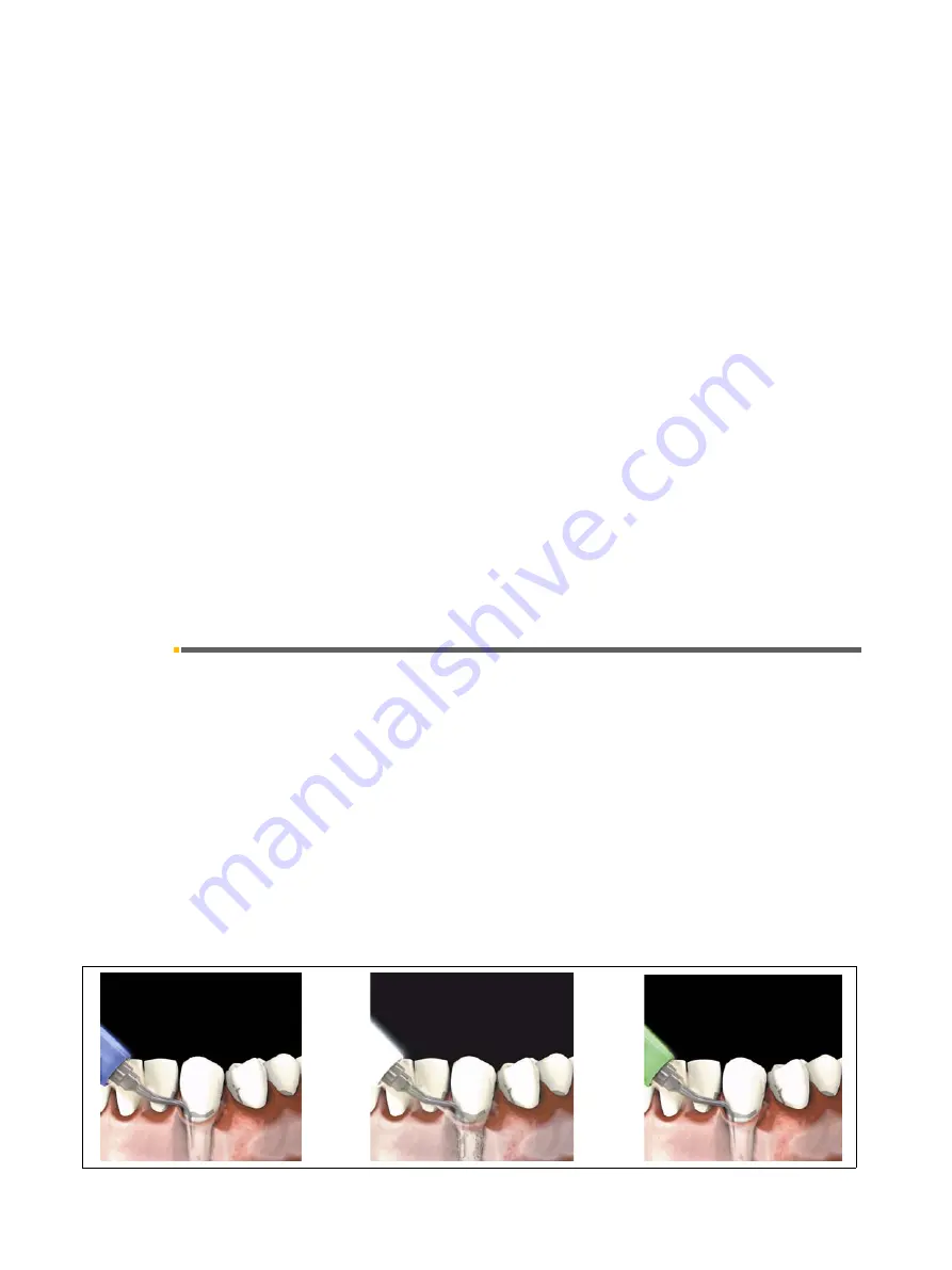
Patient communication system
A mode with three images illustrating the detection func-
tion can be invoked for communication with the patient.
You can change to the patient communication mode by
simultaneously pressing the "
Large rinsing media
tank
" and "
PERIO 2
" keys. The first image then appears
on the display.
You can then switch back and forth between the individ-
ual images with the "
PERIO 1
", "
PERIO 2
" and
"
PERIO 3
" keys.
You can quit the patient communication mode by simul-
taneously pressing the "
Large rinsing media tank
" and
"
PERIO 2
" keys once again.


