61 77 476 D 3495
D 3495
.
201.01.02
.
02
08.2007
37
Sirona Dental Systems GmbH
8
Use of the X-ray sensor
Operating Instructions and Installation
XIOS USB
Half-angle technique without radiation limiter
8.4
Half-angle technique without radiation
limiter
Depending on the size of the tooth or the location of the area to be exposed,
place the sensor in the patient’s mouth vertically or horizontally.
Here the patient can immobilize the sensor by holding it himself.
8.4.1
Endodontic exposures
Explanation
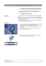
A special sensor holder is available for endodontic exposures.
z
This sensor holder is color-coded
green
.
z
The following illustrations show how to attach the sensor holder to the
hygienic protective sleeve with sensor.
Preparation
1.
Slide the sensor into the hygienic protective sleeve. When doing so, follow
the instructions for sensors.
2.
Select the green sensor holder for endodontic exposures and remove the
protective foil from the adhesive surface.
3.
Anterior tooth exposures:
To take endodontic anterior tooth exposures,
position the sensor holder on the edge of the sensor near the cable
4.
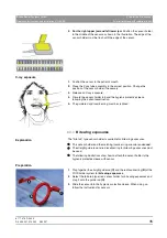
Posterior tooth exposures:
To take endodontic posterior tooth
exposures, position the sensor holder in the middle of the sensor.
X-ray exposure
1.
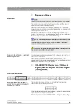
Position the sensor in the patient’s mouth.
2.
Place the X-ray tube assembly in the correct position. Change the
position of the sensor holder if necessary.
3.
Release an X-ray exposure.
4.
Dispose of the holder and the hygienic protective sleeve following the
patient examination.


